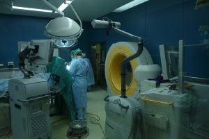
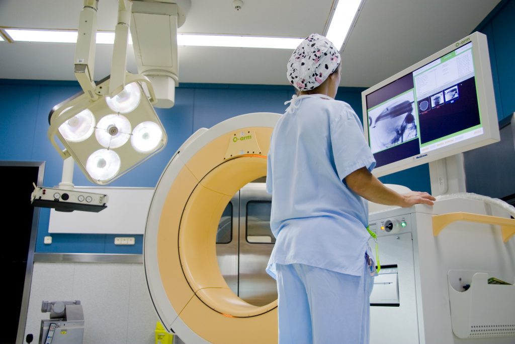
 Just like when the introduction of X-rays into operating theatres represented a huge step forward for surgical techniques, the introduction of the intraoperative CT or scanner also implied new progress in intraoperative imaging systems. The General Hospital of Catalonia had the first intraoperative CAT scan in Spain,
and so our group was the pioneer in the introduction and development of these units on a European level. This system enables precise 3D location of the surgical procedure therefore making it less aggressive for the patient. Since its introduction in operating theatres, this system has undergone enormous development, mainly driven by the growing demand placed on the system by professional physicians, who constantly demand more precise tools, as well as by the patients themselves who have more and more information about the procedures and treatments affecting them. Its use can mainly be divided into two situations:
- Update of Neuronavigation units: on the one hand enabling refreshing navigation systems at any time during the surgical procedure, minimising errors resulting from the lack of the latest navigation information.
- Verification of the location of prosthesis: it is also extremely useful for verifying the correct placement of prosthesis in orthopaedic surgery, spinal surgery or cerebral cortex stimulation systems (e.g. treatment of pain) or deep brain stimulation (DBS, surgery for Parkinson’s disease), as well as ENT surgery (sinus surgery) and by groups of maxillofacial and plastic surgeons.
The introduction of the intraoperative CAT scan provided a new tool in operating theatres to enable performing more and more precise and less aggressive surgical procedures.


 Just like when the introduction of X-rays into operating theatres represented a huge step forward for surgical techniques, the introduction of the intraoperative CT or scanner also implied new progress in intraoperative imaging systems. The General Hospital of Catalonia had the first intraoperative CAT scan in Spain,
